A new crystallographic technique developed at the University of Leeds is set to transform scientists’ ability to observe how molecules work.
A research paper, published in the journal Nature Methods, describes a new way of doing time-resolved crystallography, a method that researchers use to observe changes within the structure of molecules.
Although fast time-resolved crystallography (Laue crystallography) has previously been possible, it has required advanced instrumentation that is only available at three sites worldwide. Only a handful of proteins have been studied using the traditional technique.
The new method will allow researchers across the world to carry out dynamic crystallography and is likely to provide a major boost in areas of research that rely on understanding how molecules work, such as the development of novel smart materials or new drugs.
Understanding how structure and dynamics are linked to function is key to designing better medicines that are targeted at specific states of molecules, helping to avoid unwanted side effects.
“A time-resolved structure is a bit like having a movie for crystallographers,” said Professor Arwen Pearson, who led a team of researchers in the University's Faculty of Biological Sciences and School of Chemistry. “Life wiggles. It moves about and, to understand it, you need to be able to see how biological structures move at the atomic scale. This breakthrough allows us to do that.”
Traditional X-ray crystallography fires X-rays into crystallised molecules and creates an image that allows researchers to work out the atomic structure of the molecules. A major limitation is that the picture created is the average of all the molecules in a crystal and their motions over the time of an experiment.
Dr Briony Yorke, the lead researcher on the project, said: “A static picture is not very helpful if you want to observe how molecular structures work. It is like trying to find out how a car works without being allowed to run the engine. You can look at the spark plugs and the piston and maybe take a guess at how it is going to function, but it is hard to really understand something without seeing it in action.”
The existing method of getting around the problem could be compared to the laborious process of making an animated film. Scientists “synchronise” a set of molecules in an identical state and then activate, or “pump”, the changes in the molecules. They take a crystallographic snapshot of the structure after a set time. The researchers then have to begin the whole experiment again: synchronising the molecules, “pumping” them and then taking a snapshot a bit later in the process. Slowly, they build up a moving picture.
This pump-probe approach was first proposed in Nobel Prize winning research by the British chemist George Porter in the 1940s and has had a huge impact on our understanding of chemistry. However, a major limitation of the “pump-probe” approach for crystallographic experiments is that the snapshots are only “exposed” for a moment (often as short as 100 million millionths of a second) in order to capture the molecular movements. That means there is very little time to deliver enough light to create a crystallographic image.
There are only three “synchrotrons”, large X-ray generators, in the world—in the US, France and Japan—that are capable of delivering a powerful enough beam.
The new method uses clever maths (a Hadamard Transform) to open up the field to much less powerful synchrotron “beamlines”, advanced laboratories that scientists use to harness powerful synchrotron light for crystallography and other techniques. This will enable facilities such as the UK’s own synchrotron, Diamond Light Source, to do time-resolved crystallography.
As in Porter’s method, in the new approach researchers synchronise their molecules and activate them. However, they then make a series of crystallographic “probes” of the moving structures using a pattern of light pulses. These pulses build up a single crystallographic image—a bit like a long exposure photograph. The researchers then repeat the experiment using a different pattern of light pulses and create a different “long exposure” image. This process is repeated until all of the pulse patterns created using a mathematical formula have been completed.
Each of the “long exposure” images created from the pulse patterns is blurred, but the differences in the images and between the pulse patterns that created them allow researchers to extract a moving picture of the molecules’ changing structures.
Professor Pearson said: “The great thing about this method is that we don’t need the clear snapshots, and therefore the very strong light, required by the Porter method. This is a completely new way of doing a time-resolved experiment and overcomes many of the current limitations.”
Co-author Professor Godfrey Beddard, Emeritus Professor of Chemical Physics at the University of Leeds, said: “We demonstrate this method for crystallography, but it will work for any time-resolved experiment where the probe can be encoded. This new method means that, instead of having to go to one of the three instruments in the world that can currently do time-resolved crystallography, you can go to any beamline at any synchrotron—basically it massively opens the field for these kind of experiments.”
Co-author Dr Robin Owen, Principal Beamline Scientist at Diamond Light Source, said: “The beauty of the approach is that it uses existing equipment in a new way to facilitate new science. The novel use of the Hadamard transform, or multiple-exposure, approach helps open the door for time-resolved science at a much wider range of beamlines and synchrotron sources than is currently possible. By exploiting the approach we will be able to obtain multiple sequential images of a protein while it carries out its function, providing a much clearer understanding of the relationship between structure and function.”
Professor Paul Raithby, Chair of Inorganic Chemistry at the University of Bath, a leading expert on time-resolved crystallography, who was not one of the authors of the paper, said: “This is a very exciting development in the area of macromolecular and molecular crystallography. The new method will allow us to "watch" chemical and biological processes as they happen in a way that has not been possible previously, and open up new vistas of research into the dynamics of molecules and materials. What is particularly exciting from my point of view is that the method can be used in my area of research to look at how "smart" materials function in real time and help in the design of the next generation of electronic devices and sensors."
The research was funded by the Wellcome Trust and was conducted at the University of Leeds and the Diamond Light Source. Professor Pearson is now Professor of Experimental Biophysics at The Hamburg Centre for Ultrafast Imaging (CUI) of Universität Hamburg. Dr Yorke is now a postdoctoral research fellow, also at Universität Hamburg.
Image information:
Homepage banner image: The equipment on I04, one of Diamond Light Source's macromolecular crystallography beamlines (provided by Diamond Light Source)
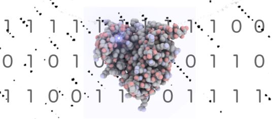
Image on this page: Thaumatin molecule against Hadamard matrix background (provided by Dr Briony Yorke)
Further information
Professor Arwen Pearson, Dr Yorke and Professor Godfrey Beddard are available for interview and can be contacted through the University of Leeds press office.
Contact: Chris Bunting, Senior Press Officer, University of Leeds; phone:+44 113 343 2049 or email c.j.bunting@leeds.ac.uk
Dr Owen can be contacted through Silvana Westbury in the Diamond Light Source press office; phone: +44 1235 778130 or email silvana.westbury@diamond.ac.uk.
The full paper: Briony A Yorke, Godfrey S Beddard, Robin L Owen, Arwen R Pearson “Time-resolved crystallography using the Hadamard Transform” is published in Nature Methods (2014) (DOI: 10.1038/nmeth.3139; URL: http://dx.doi.org/10.1038/nmeth.3139).