The healthcare company Roche has acquired the intellectual property and technology of the Leeds Virtual Microscope (LVM), an innovative system designed to help pathologists making cancer diagnoses.
The technology, developed at the University of Leeds and the Leeds Teaching Hospitals NHS Trust, is expected to be integrated into Roche’s Digital Pathology (DP) portfolio.
Professor Roy Ruddle, from the University of Leeds’ School of Computing and co-leader of the project, said: “The system is currently the only microscope in the world that provides pathologists with a field of view as large as a conventional light microscope. We also have developed a highly innovative user interface that lets pathologists interactively view large patient cases of 100 slides or more to make complex diagnoses.”
Virtual microscope systems currently on the market have typically restricted users to less than a third of the field of view of the LVM—slowing down diagnosis and deterring adoption by clinicians.
An evaluation published in 2014* found that hospital consultants with little experience of the LVM were able to process cases in almost the same time as using familiar traditional techniques.
Dr Darren Treanor, Consultant Pathologist with Leeds Teaching Hospitals NHS Trust and the University's Leeds Institute of Cancer and Pathology, said: “We are not just offering parity with glass-based diagnostics. Going digital means you are no longer tied to the glass. Second opinions are a common part of cancer diagnosis and in some cases that means the glass slide has to travel across the country or abroad to get to the right people. That can slow down the system and introduce delays for patients.”
“A major teaching hospital like St James’s University Hospital in Leeds produces in excess of 250,000 slides in a year and all that glass has to be archived. Digitising the system would not only remove the need for physical archives, but also give doctors much faster access to past cases,” Dr Treanor said.
“But most importantly, when we evaluated the LVM with our medical colleagues, they were impressed with its speedy interface, and eager to have the tool available in their laboratories. As a result, we will be deploying the LVM for diagnostic use in our hospital, starting with breast cancer in 2016.”
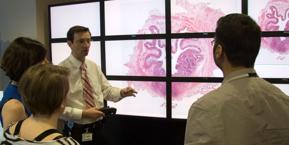
The LVM has been developed to run on systems that range from laptops, to high-definition medical displays, and 50 megapixel “Powerwalls”. At St James’s University Hospital in Leeds, the Powerwall version has been used for more than 5 years to train pathologists, with the large and highly detailed virtual slides forming the basis of small-group discussions and training courses.
In 2016 the LVM will be rolled out across 13 hospitals in Yorkshire Deanery, allowing trainee pathologists to benefit from the system and incorporate digital pathology in their learning.
The LVM project won the 2014 Yorkshire & Humber NHS Innovation Award for Medical Devices and Diagnostics and is an outstanding example of multidisciplinary research at the University, involving researchers from the Leeds Institute of Cancer and Pathology, the School of Healthcare, the School of Computing, as well as the Leeds Teaching Hospitals NHS Trust.
It received funding from the National Institute for Health Research, the Medical Technologies Innovation and Knowledge Centre based at Leeds, the Engineering and Physical Sciences Research Council, the Pathological Society of Great Britain and Ireland, the Higher Education Funding Council for England, the higher-education information technology organisation JISC and the Leeds Teaching Hospitals Charitable Trustees. The educational roll-out is funded by the Yorkshire and the Humber Deanery.
*For details of the 2014 evaluation see: http://www.virtualpathology.leeds.ac.uk/research/Publications/pub_docs/rr/randell-humanpathology-evaluation2.pdf
Image information: The Leeds Virtual Microscope displays slide on a Powerwall (©University of Leeds)
Further information
Dr Treanor and Professor Ruddle are available for interview.
Images of tissue slides and of the LVM system are available on request.
Contact: University of Leeds press office on +44 113 343 4031 or email pressoffice@leeds.ac.uk