Engineers have shown it is technically possible to guide a tiny robotic capsule inside the colon to take micro-ultrasound images.
Engineers have shown it is technically possible to guide a tiny robotic capsule inside the colon to take micro-ultrasound images.
Known as a Sonopill, the device could one day replace the need for patients to undergo an endoscopic examination, where a semi-rigid scope is passed into the bowel – an invasive procedure that can be painful.
Micro-ultrasound images also have the advantage of being better able to identify some types of cell change associated with cancer.
The Sonopill is the culmination of a decade of research by an international consortium of engineers and scientists. The results of their feasibility study are published today in the journal Science Robotics.
“The technology has the potential to change the way doctors conduct examinations of the gastrointestinal tract.”
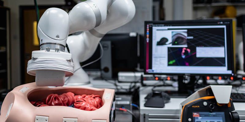
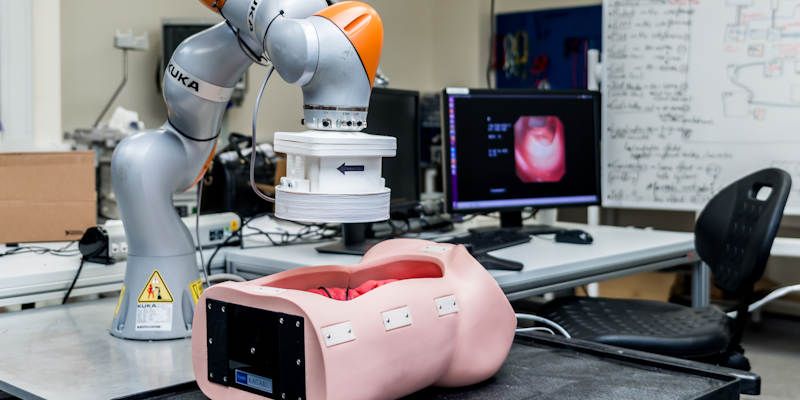
The consortium has developed a technique called intelligent magnetic manipulation. Based on the principle that magnets can attract and repel one another, a series of magnets on a robotic arm that passes over the patient interacts with a magnet inside the capsule, gently manoeuvring it through the colon.
The magnetic forces used are harmless and can pass through human tissue, doing away with the need for a physical connection between the robotic arm and the capsule.
An artificial intelligence system (AI) ensures the smooth capsule can position itself correctly against the gut wall to get the best quality micro-ultrasound images. The feasibility study also showed should the capsule get dislodged, the AI system can navigate it back to the required location.
Professor Pietro Valdastri, who holds the Chair in Robotics and Autonomous Systems in Leeds' School of Electronic and Electrical Engineering and was senior author of the paper, said: “The technology has the potential to change the way doctors conduct examinations of the gastrointestinal tract.
“Previous studies showed that micro-ultrasound was able to capture high-resolution images and visualise small lesions in the superficial layers of the gut, providing valuable information about the early signs of disease.
“With this study, we show that intelligent magnetic manipulation is an effective technique to guide a micro-ultrasound capsule to perform targeted imaging deep inside the human body.
“The platform is able to localise the position of the Sonopill at any time and adjust the external driving magnet to perform a diagnostic scan while maintaining a high quality ultrasound signal. This discovery has the potential to enable painless diagnosis via a micro-ultrasound pill in the entire gastrointestinal tract.”
Sandy Cochran, Professor of Ultrasound Materials and Systems at the University of Glasgow and lead researcher, said: “We’re really excited by the results of this feasibility study. With an increasing demand for endoscopies, it is more important than ever to be able to deliver a precise, targeted, and cost-effective treatment that is comfortable for patients.
“Today, we are one step closer to delivering that.
“We hope that in the near future, the Sonopill will be available to all patients as part of regular medical check-ups, effectively catching serious diseases at an early stage and monitoring the health of everyone’s digestive system.”
The Sonopill is a small capsule – with a diameter of 21mm and length of 39mm, which the engineers say can be scaled down. The capsule houses a micro ultrasound transducer, an LED light, camera and magnet.
A very small flexible cable is tethered to the capsule which also passes into the body via the rectum and sends ultrasound images back to a computer in the examination room.
The feasibility tests were conducted on laboratory models and in animal studies involving pigs.
Diseases of the gastrointestinal tract account for approximately 8 million deaths a year across the world, including some bowel cancers which are linked with high mortality.
The research was funded by the Engineering and Physical Sciences Research Council, the Royal Society and the US National Institutes of Health.
Further information
Contact University of Leeds Media Relations Officer on +44(0)113 3432049 or d.lewis@leeds.ac.uk.