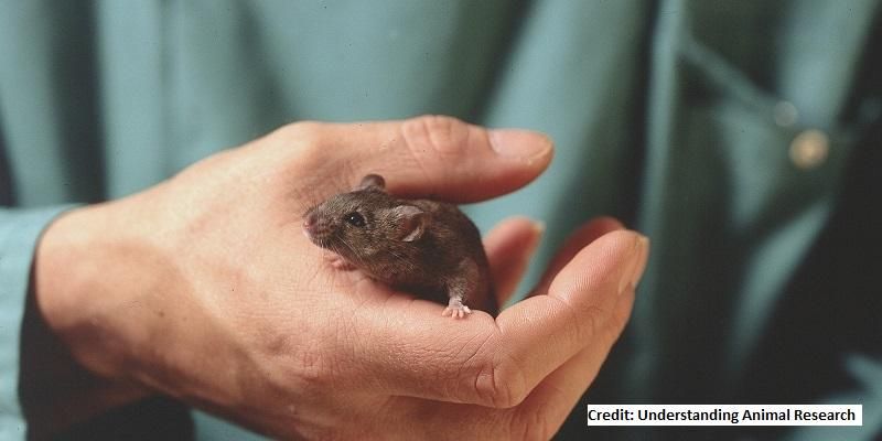
It’s two thousand times smaller and beats ten times faster – but the mouse heart holds important clues to the way heart disease develops in humans.
To better learn from the animals, the University of Leeds has opened a £6 million centre to develop new ways of studying the mouse heart using non-invasive scanning techniques.
Scientists have taken the technologies normally found in major hospitals, such as magnetic resonance imaging (MRI), and scaled them down for use with small animals.
In the human health setting, MRI scans and an associated technique called magnetic resonance spectroscopy (MRS) provide doctors with detailed information about the heart and the way it is functioning.
But because the mouse heart is so small and beats so rapidly, it is technically challenging to use existing human sized diagnostic technology to accurately measure what is happening.
Under the lead of Jurgen Schneider, Professor of Biomedical Imaging at the Leeds Institute of Cardiovascular and Metabolic Medicine, a research team is developing new ways of analysing the data that comes from the advanced scanning process.
Some of the techniques will relate to developing both the hardware and software used in the scanning.
The techniques the team have developed will create three dimensional films of hearts so scientists can look at the structure of the organ and measure the metabolism within tissue.
The animals are anaesthetised during the scanning process.
Technical challenges
Professor Schneider, who recently moved to Leeds from the University of Oxford, said: “There are huge technical issues in using established imaging technology on such small animals.
“One area we want to look at is the time it takes to run a scan and to reduce that – but we also want to develop entirely new techniques, to be able to see molecular changes that might be taking place inside the animal’s heart.”
Mouse and rat hearts are not identical to human hearts – but they are a close alternative for scientists to investigate the cause and progress of heart conditions which are the cause of major illnesses and premature death.
Alternative techniques used in animal heart research such as electrocardiogram (ECG) or echocardiography, an ultra sound scan of the heart, only give limited information. Blood flow monitoring is invasive.
Magnetic resonance imaging involves analysis of living tissue and not samples taken from a dead animal.
Professor Schneider said: “The aim of this research is to develop tools that will provide an insight into heart disease that can be fed back to the clinical environment.
“Studying how animals like rats and mice function can give us a very valuable insight into how human organs operate, opening new ways to understand and treat heart disease and other disorders.”
More humane animal research
The development of small animal scanning is a step towards the 3R principles that guide animal research – where scientists look for ways of reducing the number of animals killed in research, or replacing animals with other research techniques or refining the research methods – so they are less invasive.
The University supports the work of the National Centre for the Replacement, Refinement and Reduction of Animals used in Research, and is also a signatory to the Understanding Animal Research Concordat on Openness on Animal Research.
The British Heart Foundation contributed £1.9 million towards the cost of the imaging suite. Professor Metin Avkiran, Associate Medical Director at the British Heart Foundation, said: “The BHF is proud to have supported the construction of the new experimental imaging centre at the University of Leeds.
"This state-of-the-art building will be home to the very best imaging equipment and will help to reveal the structure and function of the heart, how things go wrong in disease, and how new treatments may work in greater detail than ever before.
“These non-invasive imaging approaches also allow studies that adhere to the principles of the 3Rs (Replacement, Reduction, Refinement) towards minimising the number of animals used in research and maximising their welfare.
“The establishment of new centre, thanks to support from the BHF, is a powerful new addition to the armoury that scientists and doctors can deploy in the fight against the devastation that is caused by heart and circulatory disease.”
Further information
Journalists with questions or interview requests should contact David Lewis in the University press office on 0113 343 8059 or email d.lewis@leeds.ac.uk.